Ernst Haeckel (1834-1919) was a German biologist-philosopher,
who worked with different scientific areas. He introduced new names for his
discoveries in different scientific fields such as the terms ecology,
anthropogeny, phylum, the kingdom
Protista, phylogeny, stem cell and also proposed theRecapitulation1
Theory.
Ernst Haeckel (1834-1919) was a German biologist and philosopher.
Images of dog and human embryos, looking almost identical at 4 weeks (most left upper panel) then differing at 6 weeks (left upper panel at right continuously). Also above at right hand side, shown a 6 week turtle embryo and below a 8 day hen embryo, presented by Haeckel in 1868 as convincing proof of evolution. The pictures of the earliest embryonic stages are now considered inaccurate (Image from Richardson et al. 1998).
The Recapitulation Theory was proposed to create a synthesis on basis of Lamarckian and
Darwinism theories. The Recapitulation
Theory says:
“the ontogeny2
recapitulates phylogeny3”, (look the image above) which it means that the whole development
process repeats or resemble the complete successive evolutionary stages,
beginning with his common ancestor passing through several organism that he had
already evolved until reach his own proper form. It’s said, when an organism is
developing, he remakes and pass through every embryonary development stage that
compound his whole evolutionary lineage-history. In other words, each of the
individual states of a species crosses throughout embryonic development,
represents one of the adult forms that appeared in their evolutionary history. For
example, Haeckel proposed that the pharyngeal grooves between the pharyngeal
arches in the neck of the human embryo resembled gill slits of fish, thus
representing an adult "fishlike" developmental stage as well as
signifying a fishlike ancestor. Ernst Haeckel made drawings of his observation
studies and there were several controversies about Haeckel drawings where he
shows the variety of embryology resembling between organisms. There was several
misunderstanding about his work at that time.
During the nineteenth century, there were several scientists
agree with Haeckel’s ideas “that all vertebrates looked very similar at an early
stage, in what was thought of as a common ideal type”. But others differ
proposing different perspectives views like Karl Ernst von Baer. He thought “the
early general forms diverged into four major groups of specialized forms
without ever resembling the adult of another species, showing affinity to an archetype (is a universally understood
symbol, term) but no relation to other types or any Transmutation of species
(theory that described the altering of one species into another)”.
Actually, the Recapitulation Theory does not
have any applicability (see figure 1)and
is until today rejected by his literacy in modern biology. For instance, if you
observe a chick's development, you will see that the chick embryo may resemble
the embryos of reptiles and fish at points in its development, but it doesn't
recapitulate the forms of its adult ancestors (figure 2). Another example is the axolotl, which
evolved from a salamander ancestor that had internal gills in the adult stage.
However, the axolotl never develops through a stage with internal gills; its
gills remain external (figure 3).
Figure 1. Representation that organisms don't stop in a intermediate state showing successive changes to acquire or resemble the anterior stage of a full adult form see in during the development (Richardson M. et al; 1998).
Figure 2. The chicken don't recapitulates his adult ancestors such as it was formulated by Ernst Haeckel.
recapitulation: to restate briefly : summarize; to repeat the principal stages or phases of something.
ontogeny: ontos means: “to be” and geny means “mode of production”; the history of structural change in a unity, which can be a cell, an organism, or a society of organisms, without the loss of the organization which allows that unity to exist.
phylogeny: is the story of the evolutionary development of a group of organisms.
References:
Embryo Project Encyclopedia. 2007. Retrieved August 2011. "In 1874 His published his Über die Bildung des Lachsembryos, an interpretation of vertebrate embryonic development. After this publication His arrived at another interpretation of the development of embryos: the concrescence theory, which claimed that at the beginning of development only the simple form of the head lies in the embryonic disk and that the axial portions of the body emerge only later."
Ernst Haeckel — Britannica Concise (biography) Encyclopædia Britannica Concise, 2006.
Evolution and Development for the 21st Century. Retrieve on Sunday, march 31 2013 from: Stephen Jay Gould http://evolution.berkeley.edu/evosite/history/modevdev.shtml
Learning about evolutionary history. Retrieve on Sunday, march 31 2013 from: http://evolution.berkeley.edu/evolibrary/article/0_0_0/evodevo_02
Michael K. Richardson. 1998. "Haeckel's embryos continued." Science 281:1289, quoted in NaturalScience.com webpage Re: Ontogeny and phylogeny.
I think it
is important to recognize scientists who made important contributions to
science, in whatever field and in whatever time they made their
discoveries.Here I would like to talk
about the significance of the discovery of the homeobox genes.Several scientists contributed significant
work to that milestone in evolutionary and developmental biology.After fertilizing an egg, cells divide
gradually until it forms tissues like skin, muscle, nerves and others.However, before cells begin to specialize,
the body begins to designate mayor regions like head, trunk and tail.The first scientist discovering the head to
tail or the determination of
the anteroposterior axis was Ross G. Harrison of
the Yale University.In 1918 Harrison
showed that when he took embryonic cells from an amphibian from a certain
region in neural stage and transplanted them to another embryo, this one formed
an extra forelimb or extremity.His
conclusion was that these cells already knew what to do in whatever place they
were. He initiated the era of cell
transplantation experiments.After World
War II scientists changed from transplantation experiments to genetic studies
of how the body took shape.In 1948
Edward B. Lewis of the California Institute of Technology started to analyze
homeotic mutations in the fruit fly Drosophila
melanogaster.By mutating homeotic
genes some body parts are replace with other structures from other body parts.Examples are the bithorax mutant that has two pairs of wings and not just one pair
as usual, or the antennapedia mutant
that has legs instead of antenna in the head.Lewis found that the mutated genes were mayor players in the body
formation.But it was not until 1980’s
that scientist could isolate specific genes so they could study them.
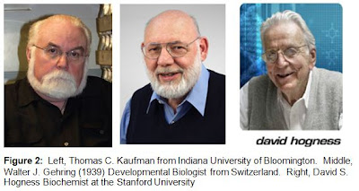
David S. Hogness and Welcome Bender of the
Stanford University were the first to isolate the genes Ultrabithorax, Abdominal A and
Abdominal B and the Bitorax complex.At the same time Walter J. Gehring and
Richard L. Garber of the University of Basel and Thomas C. Kaufman of the Indiana University isolated
genes like Antennapedia, Labial,
Proboscapedia, Deformed and Antennapedia. All this work was done in fruit flies, but
scientists became more interested in finding a similar pattern in a vertebrate
model organism.
Even there has been done significant genetic work on mice, there was no such comparable homeotic gene
found like the ones in drosophila.It
was the time when Eddy M. De Robertis, Guillermo Oliver and Christopher V. E.
Wright had this weird idea to us the
homeobox probes that McGinnis andGehring used in the fruit fly.However they had not much support from the
other scientists because at that time no one believed that two such different
species like fruit flies and frogs could have genes in common. Nevertheless they made the experiment and
found a surprise; in frog exists also a homeobox region that seems to control
vertebrate development.This is an
example how different animal models and different strategies can help revel
important discoveries in science as in this case in evo devo research.Therefore it is important to mention new knowledge;
however it is also important to mention the people behind these important
discoveries.
Reference:
E. M. De Robertis, G. Oliver, C. V. E. Wright,
1990. Homeobox genes and the vertebrate body plan. Scientific american.
Have
you ever questioned, how is possible that an organism acquires new features
based on their own genes? Many genes and signaling pathways have multiple
developmental roles. For example, the transcription factor Distalless is required to organize the development of legs, wings,
and antennae of all insects, but in some butterflies, it is also expressed
later in specific positions on the developing wing, where it is involved in
setting up the color patterns known as "eyespots" (see Figure 1).
Such cases suggest that, over the course of evolution, genes and pathways have been
redeployed to serve new functions such as in the case of reptiles scales that
through million of years become bird’s feathers (see figure 2 and Youtube video). Change in
the function of pre-existing features in adaptive evolution has been known ever
since Darwin. Gould and Vrba (1982) coined the term exaptations to refer to novel uses of pre-existing morphological
traits. Developmental
biologists have used the terms recruitment
(Wilkins 2002) and co-option
(True and Carroll 2002) to refer to the evolution of novel
functions for pre-existing genes and developmental pathways.
Figure 1. Co-option of developmental circuits in the evolution of novelties. (A) Butterfly "eyespots" are the developmental products not only of genes for pigmentation, but also of many co-opted genes and pathways that play important roles in establishing the body plan. (B) Co-option of the vertebrate Hoxa genes during evolution of the tetrapod appendage. Ancestrally, Hox genes were expressed only along the anterior-posterior axis of the developing body. The evolution of paired fore-and hindlimbs involved novel gene expression, presumably using novel enhancer sequences of Hoxa 9-13.
Figure 2. Wallace’s flying frog (Rhacophorus nigropalmatus), an inhabitant of the rain forest canopy in southeastern Asia, glides from tree to tree with the aid of its toe webbing, which is much more extensive than in most other tree frog species. (Photo by Stephen Dalton/Minden Pictures, Inc.)
The Origin of the brain
Feather Evolution
Co-option of single genes
for new functions may be common. The members of many gene families have
diversified into different developmental and physiological roles. In one of the
most interesting such cases, the diverse crystals in proteins of animal eye
lenses have been co-opted from a number of genes (Figure 3). Two types of
crystallins, α. and β, which are common to all vertebrates, are derived from
stress proteins, which have help stabilize cellular functioning during environmental
stresses, such as excess heat. Various lineages of animals have also derived
taxon-specific crystalline from distinct enzymes, such as lactate dehydrogenase
in reptiles and glutathione-S-transferase in cephalopods. In the eye lens,
crystallins are expressed at high levels and packed tightly into transparent
matrices that are resistant to environmental stress and are designed to endure
for the entire adult life of the animal. To achieve this function, many
crystallins have undergone amino acid substitutions since they were co-opted
from their ancestral function, but in most cases these proteins still share
extensive homology with the ancestral proteins. These crystallins are derived
from duplicated enzyme genes. In other cases, gene duplication has not
occurred, and both the crystalline and the enzyme are encoded by the same gene
(e.g., τ crystallin: α enolase in fishes, reptiles, and birds).
Figure 3. Two modes by which variation in the expression of a developmental regulatory gene may be co-opted to promote novel morphogenetic features. (A) Temporal co-option. Gene expression that persists after the critical period may be utilized during evolution to regulate novel morphogenetic processes. (B) Spatial co-option. This novel expression may be co-opted to promote morphogenesis of an ancestral feature in the novel region of the body.
References:
Bock, W.J. (2000). "Explanatory History of the Origin of Feathers". Amer. Zool. 40 (4): 478–485.
Wistow, G., and J. Piatigorsky, J. (1988). Lens crystallins: The evolution and expression of proteins for a highly specialized tissue. Annu. Rev. Biochem. 57: 479-504.
Dr.
Antonia Monteiro (Assistant Professor in Yale University since
2006; see image 1). One
of her interest is the study of the origins of wing butterflies eyespot (image 2). Based on the theory of co-option(or exaptation, read blog# 13 for more information), that explains that genes
possibly shift in the function of a trait during evolution. For example, a
trait can evolve because on a time can served for a particular function, but
subsequently it may come to serve another function.
Image 1. At left side Omar Delannoy-Bruno, student in developmental biology field; (at center) Dr. Antonia Monteiro (main guest invited); (at right side) Rey J. Rosa Morales (author of this entry blog) in Old San Juan, Puerto Rico.
Image 2. Eyespot in butterfly wing
According
to Monteiro and her group (2012) they discover that from both morphological and
developmental perspectives of homology [1,3,4], nymphalid eyespots and an associated
gene cluster arose a single time, early in the evolution of the Nymphalidae
(see image 3). In addition, this single origin, multiple losses of gene
expression have occurred, suggesting a novel means in which complex traits
originate: from an initial gene regulatory network co-option followed by
stream-lining of extraneous network elements. Moreover, they also found that
the origin of eyespots was concurrent with the origin of the gene expression
patterns, approximately 90 million years ago. This finding suggests that
complex traits such as butterfly eyespots may initially evolve by re-deploying
pre-existing gene regulatory networks, which are subsequently trimmed of genes
that are unnecessary in the novel context.
Image 3.Nymphalidae subfamilies
In the study, they obtain the results from a morphological
assessment of homology; they used Mayr’s definition where ‘‘a feature is
homologous in two or more taxa if it can be traced back to the same feature in
the presumptive common ancestor.’’ [2]. If eyespots are homologous, there
should be a single origin of this trait; in contrast, multiple origins of
eyespots within the Nymphalidae would demonstrate that the traits are not
homologous [7]. (see Figure 1 and 2).
Figure 1. Origins of eyespots and associated gene expression. (A) Origin of eyespots inferred from 399 nymphalid and 29 outgroup species from phylogeny in reference 3. (B) Origin of expression in eyespot centers inferred from gene expression profiles of 23 species. Presence or absence of expression of genes in future eyespot centers indicated by black and white boxes, respectively, and grey boxes indicate species/gene combinations for which expression data are unavailable. Green bars indicate two independent origins of eyespot-associated Antp expression. In both (A) and (B), divergence times (in millions of years) are from reference 3, 7; red bars on the phylogeny indicate the possible locations of the single origin of eyespots, while gold bars indicate possible locations for the single origin of gene expression for sal, Notch, Dll, and possibly en in the eyespot centers. Asterisks (*) indicate species for which expression data are from [5,6].
Figure 2. Regulatory network simplification in a complex trait. Following the origin of a complex trait and its underlying developmental gene regulatory network, genes that are non-functional or unnecessary may be subsequently removed from the network (genes 2 and 3), without eliminating the trait. Genes expressed in homologous traits of all taxa may represent a ‘core network’ of regulatory elements (genes 1 and 4) that are necessary for the development of the novel trait.
References:
Abouheif
A (1999) Establishing homology criteria for regulatory gene networks: prospects
and challenges. In: Bock GR, Cardew G, editors. Homology. Novartis Foundation
Symposium 222. Chichester: Wiley. pp. 207–225.
Mayr
E (1982) The Growth of Biological Thought. Cambridge, MA: Harvard Univ Press.
Monteiro
A (2012) Gene regulatory networks reused to build novel traits: Co- option of
an eye-related gene regulatory network in eye-like organs and red wing patches
on insect wings is suggested by optix expression. Bioessays 34: 181–186.
Saenko
SV, Marialva MSP, Beldade P (2011) Involvement of the conserved Hox gene
Antennapedia in the development and evolution of a novel trait. EvoDevo 2: 9.
Shirai
LT, Saenko SV, Keller RA, Jeronimo MA, Brakefield PM, et al. (2012)
Evolutionary history of the recruitment of conserved developmental genes in
association to the formation and diversification of a novel trait BMC Evol Biol
12: 21.
Wake DB, Wake MH, Specht CD (2011) Homoplasy:
from detecting pattern to determining process and mechanism of evolution.
Science 331:1032–1035.


